Types of Scans
If you or a loved one is dealing with secondary (metastatic) breast cancer, you're not alone. This page is here to provide you with clear and understandable information about the types of scans involved in managing secondary breast cancer. By understanding the types of scans you may be offered, you'll be better equipped to make informed decisions with your healthcare team and know what to expect.
Let's explore these important tools together.
On this page you will find:
Mammogram
Anyone registered with a GP as female will be invited via a letter in the post for NHS breast screening, which uses a mammogram, every 3 years between the ages of 50 and 71.
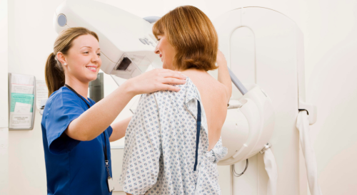
A mammogram is an X-ray that looks at breast tissue. During breast screening you'll have 4 breast X-rays (i.e., four mammograms), two for each breast.
A mammogram is one of the best ways to spot cancer that is too small to see or feel. Finding breast cancer early via a mammogram can make it more likely that initial treatment will be successful and less likely that a breast will be removed via a mastectomy.
A mammogram may be done in a breast screening unit, which could be within a hospital, or within a mobile breast screening unit which can be located in various locations in the community. The whole appointment should take around 30 minutes.
During a mammogram, you will need to undress to the waist. The breast is placed in between the mammogram machine and a plastic plate which firmly flattens the breast. This can be uncomfortable, but is necessary to obtain a clear picture, the pain will normally resolve once the procedure is finished. The procedure only takes a few moments. Two X-rays are taken, one from the side and one from above and this process is repeated for the other breast.
After your breasts have been X-rayed, the mammogram will be checked for any abnormalities. If you had a mammogram as part of routine screening, the results will be sent to you and your GP within 2 weeks of the appointment. If you have had a mammogram as part of a breast clinic appointment, the consultant will often give results then or at your next clinic appointment.
There are some risks associated with breast screening, but the medical consensus is that the benefits of a mammogram outweigh these possible risks.
The main risk is that if a mammogram diagnoses breast cancer, treatment will always be offered. However, doctors do not always know if this breast cancer will go on to be life-threatening and so you may be treated for non-life threatening cancer. Sometimes, mammograms might not identify breast cancer that is present, which is why continual self monitoring for breast cancer is important. Having a mammogram every 3 years for 20 years also very slightly increases the chances of developing cancer over a lifetime because of the X-ray exposure.
Read about Louise’s experience of a mammogram here.
CT Scan
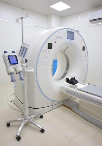 A CT (computerised tomography) scan is a medical test that uses X-rays to take detailed pictures of the inside of your body. During the scan, you'll lie on a table that moves into a big polo mint-shaped machine.
Before a CT scan, it's a good idea to wear comfortable, loose-fitting clothing, but you might need to change into a hospital gown depending on the area being scanned. You'll be asked to remove any metal objects, like jewellery or belts, as they can interfere with the quality of images. The process itself is painless and usually not uncomfortable, but you'll need to lie still during the scan.
If you are anxious about having a CT scan or have a fear of small spaces (claustrophobia) talk to a GP or a doctor at the hospital as they may be able to give you medication that can help you relax. You can also ask if you can bring someone with you to the CT scan appointment to support you.
There are two types of CT scans: (1) with contrast; (2) without contrast. Sometimes, doctors may need to inject a contrast dye to enhance the images. This can be given through a vein, rectally or in a special drink. If given through the vein your body might feel hot and flushed or you might have a metallic taste in the mouth for a short time. These feelings will usually pass quickly.
You do not need to fast if you have a CT scan without contrast. However, if the scan uses a contrast agent, you should not eat 3 hours beforehand, or as requested by your medical team. A normal CT scan usually takes less than 20 minutes. The radiographer may instruct you during the procedure and ask you to hold your breath a few times for approximately 20 seconds.
If you are having the CT scan as an outpatient you will be able to go home soon after having the scan. If you were given the contrast medium, you will be asked to wait in the hospital for around 30 minutes before leaving to make sure you don’t have a delayed reaction to the contrast agent. In some rare cases people can have an allergic reaction to the contrast medium causing dizziness, sweating, weakness and difficulty in breathing.
It usually takes between 1-2 weeks to get the results of a CT scan as the images need to be analysed by a radiologist. The radiologist will write to the doctor who made the referral for you to have a CT scan and detail the results to them. The doctor should then talk to you about your results and explain what happens next.
If you have lots of CT scans, there is a very small chance that the radiation exposure from the X-rays could elevate the risk of cancer developing. However, oncologists and radiologists will weigh up the risks of X-ray exposure against the insights the scan can provide and discuss this with you before the scan takes place.
Read about Danielle's experience of a CT scan here.
MRI Scan
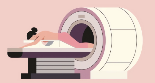
Like the CT scan, an MRI (magnetic resonance imaging) scan is also a safe and painless method to take images of the inside of the body. An MRI uses strong magnets and radio waves to create these images.
Compared with the CT scan an MRI scan is more sensitive to metal, and the presence of metal during the scanning process will be very dangerous to the machine and you. Hence, MRI may not be suitable for you if you have a metal object, such as a pacemaker or metal plates inside your body.
You will be asked to remove any jewellery containing metal, and take off any clothing with metal zippers, as well as underwire bras. You will be given a loose gown to wear. In most cases, you do not need to fast.
The process of an MRI scan is like the CT scan, but can take longer and last around 15 to 90 minutes. You will lie on a flat bed that moves into a large polo mint-shaped machine. During the scan the machine can make loud tapping noises. You may be given headphones to reduce the noise. The radiographer will speak through an intercom to give instructions and to check how you are feeling. You will be instructed to remain as still as possible during the MRI scan and you may be asked to hold your breath for a few seconds at a time. It usually takes 1 to 2 weeks to get the results of an MRI scan, unless they are needed urgently, as the radiologist who conducted the scan will need to send a report to the doctor who requested the scan.
No evidence has been found to suggest that the magnetic fields and radio waves used in an MRI scan pose a risk to the human body.
You can read about Susan’s experience of an MRI scan here.
PET-CT Scan
A PET (positron emission tomography) scan produces detailed 3D images of the inside of the body and is often combined with a CT scan to provide even more detailed images. This is known as a PET-CT scan.

Before a PET scan a small amount of a radioactive substance, known as a radiotracer, will be injected into a vein in your arm. Typically, a radiotracer that is similar to a naturally occurring sugar is used and your body will metabolise this in a similar way. The PET scan will detect the radiation given off by the radiotracer and by analysing areas where the radiotracer accumulates it's possible to learn more about how these areas of the body are working.
Cancer cells tend to use sugar at a much faster rate than normal cells resulting in the accumulation of the radiotracer enabling doctors to determine how far a cancer has spread and how well it is responding to treatment.
Typically, you will be advised to not eat anything for 6 hours before the PET-CT scan, but you can drink water. You should also avoid strenuous exercise for 24 hours before the appointment. Like in the CT scan, you will need to remove any jewellery and clothes that have metal parts, such as zips and bras with an underwire because metal can impact image quality. You can go to the toilet before having the scan.
The radiotracer will be injected into a vein in your arm around one hour before the scan because it takes some time for the radiotracer to accumulate in the cells in your body for imaging. You will be advised to relax, avoid talking and keep as still as possible whilst you wait as moving around and speaking can influence where the radiotracer goes in your body.
You will lie on a flat bed that passes into a large polo mint-shaped machine that will conduct the scan. A PET-CT scan usually lasts 30-60 minutes and is completely painless. You will be asked to remain still during the scan.
You will typically have to wait 1 to 2 weeks to get the results of a PET-CT scan as the radiologist who conducted the scan will need to make a report to send to the doctor that requested the PET-CT scan.
You shouldn't experience any side effects from the radiotracer or the PET-CT scan and will typically go home afterwards.
As a precaution, you might be advised to avoid close contact with pregnant women, babies and young children a few hours after the PET-CT scan because you'll be slightly radioactive from the radiotracer during this time. However, the radiotracer quickly becomes less radioactive over time and will naturally pass out of your body within a few hours.
Any exposure to radiation carries a risk of elevating the chances of developing cancer. However, for a PET-CT scan this is regarded as a very low risk.
X-Ray
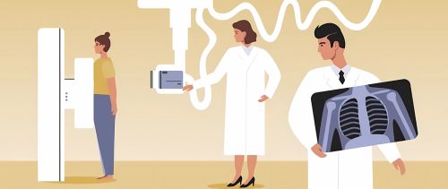
X-rays are a type of electromagnetic radiation that can be used to produce images of the inside of the body. X-rays can't be seen by the naked eye and you don't feel them. X-rays are mainly used to look at bones and joints, but they can be used to make images of soft issues, such as internal organs.
You don't usually need to do anything special beforehand to prepare for an X-ray and you can eat and drink as normal. However, it would be best to avoid wearing jewellery and clothes containing metal because these will need to be removed before the X-ray.
Unless it's an emergency, X-rays are not recommended for pregnant women.
X-rays only take a few minutes to complete and you will be able to return home afterwards. You might be told the results of the X-ray on the same day or the radiologist who conducted the X-ray will send a report to the doctor who requested the X-ray and they'll discuss the results with you shortly afterwards.
As X-rays are a form of radiation there is a risk of developing cancer from their exposure. However, the amount of radiation someone is exposed to during an X-ray is equivalent to between a few days and a few years of exposure to natural radiation from the environment. Therefore, the risk of developing cancer from an X-ray is thought to be very small and the benefits and risks of having an X-ray will be weighed up by doctors before they are recommended to you.
Ultrasound
 An ultrasound scan uses high frequency sound waves, which we are unable to hear, to generate images of the inside of the body.
You may be asked to follow specific instructions before an ultrasound scan to improve image quality. You may be advised to drink water and not go to the toilet until after the scan. You could also be advised to avoid eating or drinking for several hours before the scan. The healthcare professional organising the ultrasound will inform you of these instructions beforehand.
There are three main types of ultrasound: an external ultrasound, where the probe is moved over the skin, an internal ultrasound, where the probe is inserted into the body and an endoscopic ultrasound, where the probe is attached to an endoscope (a long, thin flexible tube) that is passed deeper inside the body.
An ultrasound typically lasts for 15 to 45 minutes and in most cases you will be able to go home soon after the scan is finished. With endoscopic ultrasounds, as sedatives can be used, you may need to stay longer at the hospital for this medication to wear off.
You could be told the results of your ultrasound straight away, but in most cases the images will be analysed afterwards and a report will be sent to the doctor who requested the scan and they'll discuss the results with you either a few days later or at your next appointment.
There are no known risks from the sound waves used in an ultrasound scan.
External and internal ultrasounds are generally painless and don't have side effects associated with them.
As endoscopic ultrasounds are more invasive they can be uncomfortable and can cause some temporary side effects such as sore throats or bloating. In some very rare cases there is also a small risk of more serious complications such as internal bleeding.
You can read about a patient's experience of an ultrasound here.
Bone Density Scan (DEXA scan)
 DEXA stands for dual energy x-ray absorptiometry. A DEXA scan uses low dose x-rays to investigate the density and therefore the strength of your bones.
DEXA scans are used if the cancer treatment you are currently taking, or have taken in the past could cause your bones to get thinner and less dense. The lower your bone density the higher your risk of breaking a bone. The progressive weakening of bones overtime is known as osteoporosis.
Breast cancer that has spread to the bones (bone metastasis) can also increase the risk of osteoporosis.
You do not need to do anything before a DEXA scan and can eat and drink as normal. However, it would be best to avoid wearing jewellery and clothes containing metal because these will need to be removed.
Unless it's an exceptional circumstance, because of the low dose X-ray exposure, DEXA scans are not recommended for pregnant women.
A DEXA scan is quick and painless and you won't feel anything. You will lie flat on a bed during the scan. You will be able to return home afterwards. The whole procedure of the DEXA scan typically takes 20 to 30 minutes.
You should get the results of the DEXA scan 1 to 2 weeks afterwards.
DEXA scans use much lower levels of radiation than standard x-rays and are typically regarded as a very safe procedure.
You can read about a patient's experience of a DEXA scan here.
Bronchoscopy
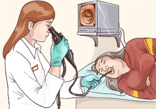 A bronchoscopy is a test to look inside the airways of your lungs. You might have a bronchoscopy so that doctors can check the cause of symptoms, further investigate any areas that look abnormal on an x-ray or scan or to take samples of cells for further tests (a biopsy).
During a bronchoscopy a thin, flexible tube will be placed down your windpipe and into the airways of your lungs. This tube will either go down your nostril or into your mouth. A nose or mouthguard will be put into place. The tip of the tube has a light and eye piece so that doctors can visualise the airways on a screen.
You will normally have a bronchoscopy as an outpatient in an endoscopy unit of a hospital. You will have a local anaesthetic with or without a sedative to help you relax. If you're having biopsy samples taken general anaesthetic will be used.
You will be given written instructions before the bronchoscopy on how to prepare. You will be asked to not eat or drink a few hours before the procedure.
A bronchoscopy takes around 20 minutes, but can take longer if biopsy samples are being collected.
You can usually go home after a bronchoscopy. Afterwards, because of the anaesthetic you will feel sleepy and a nurse will monitor you in the recovery area until you are awake. You won't be able to eat or drink anything until the anaesthetic wears off as your throat will be too numb to swallow safely. This will pass after around one hour. Someone should collect you from the hospital and stay with you overnight if you have had a sedative or anaesthetic. You should also not drink alcohol, drive or operate heavy machinery for 24 hours after having a sedative or anaesthetic.
A bronchoscopy is a safe procedure and the risks will be weighed against the benefits beforehand. A bronchoscopy can be uncomfortable and you can have a sore throat for a couple of days. There is also a risk of getting a chest infection and you should contact your doctor if you start feeling breathless and develop symptoms of an infection. You might see a small amount of blood in your spit after a bronchoscopy and should contact your doctor if this doesn't go away or gets worse as in some cases a bronchoscopy can cause internal bleeding.
You should get the results of your bronchoscopy 1 - 2 weeks after the test.
Written by Linc Yuan, University of Edinburgh
Reviewed by Senior Research Nurse Catherine Graham, Raigmore Hospital, Inverness
Date of last update: January 2024